흉선, Thymus

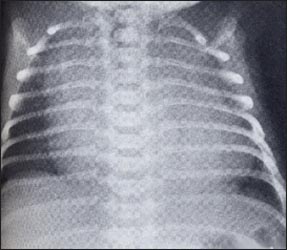
사진 1-15. 신생아 가슴 X-선 사진(정상)
가슴 X-선 사진에서 흉선이 하얗고 크게 보인다. 흉선이 흉강의 맨 위 앞 부위 대부분을 차지하고 있다. 여기서 심장의 영상과 흉선의 영상을 확실히 분별하기가 어렵다.
출처;Pediatric X-Ray Diagnosis, John Caffey, 6th Edition

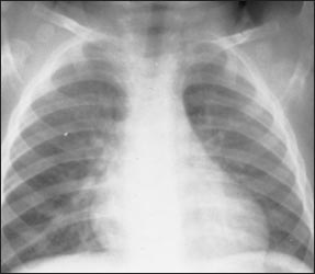
사진 1-16. 가슴 X-선 사진에서 하얗게 크게 보이던 흉선이 더 이상 보이지 않는다. 사진 1-15와 비교할 때 가슴 X-선 사진에 차이가 많은 것을 알 수 있다.
Copyright ⓒ 2011 John Sangwon Lee, MD., FAAP

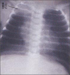
사진 1-17. 신생아 가슴 X-선사진에 나타난 흉선
이 경우 폐렴과 감별 진단해야 한다.
Copyright ⓒ 2011 John Sangwon Lee, MD., FAAP
- 흉선은 흉강의 맨 위 앞쪽, 흉골 바로 뒤쪽, 기관 앞쪽에 위치하고 있다.
- 갓 태어나서부터 첫돌까지의 흉선의 크기는 신생아의 몸의 크기나 영아의 몸의 크기에 비례해 상당히 크지만 사춘기에 이르면 흉선이 크기가 많이 위축되어 아주 적고 성인기에서는 더 위축되서 가슴 X 선상에 나타나지 않는 것이 정상이다.
- 흉선은 편도선이나 아데노이드와 같이 림프 조직의 일종이다.
- 흉선은 내분비선계에 속하기도 한다.
- 영유아의 흉선의 평균 무게는 12~15g 정도이다.
- 아주 클 때 흉선 크기는 40~60g 정도이다.
- 흉선은 항체를 생성하고, 박테리아를 죽이고, 이물을 제거하는 등 감염병이 생기지 않게 방어 역할도 한다.
- 흉선의 크기와 모양은 나이, 건강상태, 영양상태, 스트레스, 코티코스테로이드 혈 중 농도, 열, 질병, 피로 등에 따라 달라질 수 있다.
- 스트레스를 받거나 코티코스테로이드제 섭취하거나, 열, 질병, 피로 등으로 일시적으로 적어질 수 있고 그 요인이 없어지면 정상 크기로 되돌아가는 것이 정상이다.
- 신생아에게 어떤 병이 생기거나 신생아가 스트레스를 받으면 흉선 크기가 줄어들고 병이 낫고 스트레스가 없어지면 다시 보통 크기로 뒤돌아 간다. 이런 현상을 X-선 검사로 증명할 수 있다.
- 신생아때나 초기 영아기의 전후면 가슴 X-선 사진에는 흉선 영상이 크게 나타날 수 있고 폐의 위 부분과 심장의 상 부분의 영상을 가릴 수 있다. 같은 신생아나 영아의 측면 가슴 X-선 사진에는 흉선 영상이 거의 나타나지 않고 폐와 심장의 영상을 가리지 않는다. 이런 방법으로 흉선이 비정상적으로 커져있나 알아볼 수 있다.
- 선천적으로 흉선이 생기지 않아 즉 무흉선 일 때 생기는 병을 디죠오지 증후군(DiGeorge’s syndrom)이라 한다. 자가 면역성 용혈성 빈혈, 근 무력증, 소아 류마토이드 관절염(연소성 류마토이드 관절염), 갑상선염 등은 흉선 기능 이상과 관련된 질병이다.
|
다음은 “태어난 지 만 3개월이 되는 남자아이입니다. 흉선이 크다고 하는데요”에 관한 인터넷 소아청소년 건강상담 질의응답의 예 입니다. |
Q&A. 태어난 지 만 3개월이 되는 남자아이입니다. 흉선이
크다고 하는데요.
Q.
배에 가스가 차서 엑스레이를 찍었는데 폐사진이 같이 보여졌습니다. 오른쪽 폐 윗부분 반 정도가 하얗게 나와서 폐렴을 의심하여 정밀초음파를 찍었으나 폐렴은 전혀 아니고 흉선이 비대하다는 얘기를 들었습니다. 흉선은 며칠 내 또는 몇 주나 몇 달 내에 작아지니 너무 걱정 말라고는 하는데요, 흉선에 대해 알아보니 성인에서 걸리는 중증근무력증이란 병을 가진 사람이 흉선이 크다고 하구요, 흉선이 크면 기관이나 심장이나 폐를 압박해 호흡곤란 등의 문제가 있다고도 하구요.
저희아이는 특별히 호흡곤란이나 청색증을 보인적은 없는데
흉선이 크기는 하지만 기관이나 왼쪽 폐나 심장부위를 덮고 있지 않고 오른쪽 폐 윗부분으로 약간 바깥쪽으로 덮고 있어서 그런 건가요? 이런 경우는 작아지기만을 기다리면 되나요? 흉선이 커서 아이가 힘들어할 증상은 어떤 것이 있는지요? 그리고 이렇게 또래아기에 비해 흉선이 크면 나중에 커서 중증근무력증에 걸릴 확률이 더 높은지요?
그리고 흉선은 아기 때 생겨서 점차 커져서 사춘기에 제일 커지고 그 후 퇴화한다고 읽었는데요, 우리 아기는 지금 이렇게 큰데 더 커지면 어쩌지요? 진료 시 의사선생님이 작아질 거라고 하셨는데 사춘기 때까지 점차 커진다는 제가 읽은 문구와 어떻게 해석해야 하는지요?
A.
김삿갓님
안녕하세요.
질문해 주셔 감사합니다.
자녀의 나이, 성별, 과거와 가족의 병력, 진찰소견, 임상검사 결과 등 많은 정보가 있으면 더 좋은 답변을 드릴 수 있습니다. 주신 정보를 참작해 답변을 드립니다.
좋은 질문입니다.
흉선에 관해서는 P.00 흉선을 참고하시기 바랍니다.
질문하신 대로 흉선에 관해서 의료인들도 잘 모르는 점이 특히, 과거에는 많이 있었습니다.
흉선은 나이에 따라 크기와 모양과 위치 등이 다르고 열, 질병, 스트레스, 약물 등에 따라 크기가 다를 수 있습니다.
이런 이유로 같은 영아의 가슴 X-선 사진 상에 흉선이 크게 보일 때도 있고 더 작게 보일 때도 있습니다.
일반적으로 영유아들의 가슴 X-선 사진에 흉선이 보이면 건강하다는 것을 의미할 수 있습니다.
흉선은 림프 조직의 일종이고 내분비선 계통에도 속합니다.
흉선은 항체를 만들어내는 역할도 합니다.
흉선에 종양이 생길 수 있고 그 종양으로 근무력증이 생길 수 있습니다.
근무력증은 흉선에 종양이 없이도 생길 수 있습니다.
소아청소년과에서 신생아나 영아의 어떤 병을 진단하기 위해 가슴 전후면 X-선 사진을 찍을 때 흉선이 흉강의 맨 위 앞쪽이 있는 것을 볼 수 있습니다. 이 경우 가슴 측면 X-선 사진에는 흉선 영상이 거의 보이지 않을 수 있습니다.
이런 것은 다른 검사를 할 필요 없이 흉선이 정상이라고 진단 붙일 수있습니다.
신생아기에서 사춘기까지 흉선이 있는 것이 정상이고 흉선이 없으면 면역체계에 이상이 생깁니다.
흉선 종양 등의 증상 징후가 없이 흉선이 가슴 X-선 사진에 보인다고 해서 흉선에 어떤 이상 이 있다고 걱정할 필요가 없습니다.
참고로 몇 10년 전에는 흉선이 비정상적으로 크다고 방사선으로 흉선을 적게 치료를 했었습니다. 그런 치료는 잘못된 의술이었습니다.
소아청소년과에서 필요에 따라 상담하시기 바랍니다.
Google에 직접 들어가 검색하시면 필요한 정보를 영어로 더 얻을 수 있습니다. 질문이 더 있으면 또 방문하세요. 감사합니다. 이상원 드림
Thymus

Photo 1-15. Newborn chest x-ray (normal) The chest X-ray shows the thymus gland white and large. The thymus occupies most of the upper anterior portion of the thoracic cavity. Here, it is difficult to clearly distinguish the image of the heart from the image of the thymus. Source; Pediatric X-Ray Diagnosis, John Caffey, 6th Edition

Photo 1-16. The large white thymus on the chest X-ray is no longer visible. It can be seen that there are many differences in the chest X-ray when compared with pictures 1-15. Copyright ⓒ 2011 John Sangwon Lee, MD., FAAP

Photo 1-17. Thymus showed on newborn chest x-ray In this case, it should be differentially diagnosed with pneumonia. Copyright ⓒ 2011 John Sangwon Lee, MD., FAAP • The thymus is located in front of the top of the chest cavity, just behind the sternum, and in front of the trachea.
• The size of the thymus from birth to the first year of life is quite large in proportion to the size of the newborn’s body or the size of the infant’s body. It is normal not to
• Thymus is a type of lymphoid tissue like tonsils and adenoids.
• The thymus is also part of the endocrine system. • The average weight of the thymus in infants and young children is about 12 to 15 g.
• When very large, the size of the thymus is about 40 to 60 g.
• The thymus also acts as a defense against infectious diseases by producing antibodies, killing bacteria, and removing foreign bodies.
• The size and shape of the thymus may vary depending on age, health, nutritional status, stress, corticosteroid concentration, fever, illness, and fatigue.
• It may decrease temporarily due to stress, taking corticosteroids, fever, illness, fatigue, etc.
• When the newborn develops some disease or when the newborn is stressed, the thymus size decreases and returns to normal size when the disease is cured and the stress is relieved. This phenomenon can be proved by X-ray examination.
• Anterior and posterior chest X-rays of newborns or early infancy may show large thymus images and may obscure images of upper lungs and upper heart. Lateral chest X-rays of the same newborn or infant show little thymus images and do not obscure images of the lungs and heart. In this way, it is possible to detect whether the thymus gland is abnormally enlarged.
DiGeorge’s syndrome is a disease that occurs when there is no congenital thymus, that is, athymia. Autoimmune hemolytic anemia, myasthenia gravis, juvenile rheumatoid arthritis (juvenile rheumatoid arthritis), and thyroiditis are diseases associated with thymus dysfunction.
Next is “A boy who is 3 months old.
This is an example of a Q&A for children and adolescents on the Internet about “they say the thymus is large.”
Q&A. He is a boy who is 3 months old. thymus They say it’s big.
Q. I had gas in my stomach, so I took an X-ray and a picture of my lungs was shown. About half of the upper part of the right lung came out white, so I suspected pneumonia, so I took a precision ultrasound. They tell me not to worry too much because the thymus will get smaller within a few days or weeks or months. When I found out about the thymus, people with a disease called myasthenia gravis in adults say that the thymus is large.
There are also problems such as shortness of breath.
My child never showed any particular dyspnea or cyanosis. Is it because the thymus is large, but it’s not covering the trachea or the left lung or heart, but rather the upper part of the right lung slightly outward? In this case, can I just wait for it to get smaller? What are the symptoms of an enlarged thymus in a child? And if the thymus gland is larger than that of a baby of the same age, is there a higher chance of getting myasthenia gravis when they grow up later? And I’ve read that the thymus gland is born in babies and gradually enlarges, peaks in puberty and then degenerates. My baby is this big now, but what if it gets bigger? At the time of treatment, the doctor said that it would become smaller, but how should I interpret it with the phrase I read that it gradually grows until puberty?
A. Kim Satgat Hello. Thank you for asking a question. We can give you a better answer if you have a lot of information such as your child’s age, gender, past and family medical history, examination findings, and clinical test results. We will respond based on the information you have provided. That’s a good question. For thymus, see P.00 Thymus.
As you asked, there are a lot of things that doctors don’t know about the thymus, especially in the past. The thymus gland varies in size, shape, and location depending on age, and may vary in size depending on heat, disease, stress, and medications.
For this reason, sometimes the thymus may appear larger or smaller on a chest X-ray of the same infant. In general, seeing a thymus on a chest x-ray in infants and young children can mean they are healthy. The thymus is a type of lymphoid tissue and also belongs to the endocrine system. The thymus also makes antibodies. A tumor can form in the thymus, which can cause myasthenia gravis. Myasthenia gravis can develop without a tumor in the thymus.
When a pediatrician takes an anterior and posterior chest x-ray to diagnose some disease in a newborn or infant, the thymus may be seen in the upper anterior part of the chest cavity. In this case, the thymus image may be barely visible on the chest X-ray. This can be diagnosed as normal thymus without the need for other tests. It is normal to have a thymus from newborn to puberty, and the absence of a thymus causes abnormalities in the immune system. If you can see the thymus on a chest X-ray without any symptoms, such as a thymic tumor, you don’t need to worry about something wrong with your thymus.
For reference, a few decades ago, the thymus was treated with less radiation because the thymus was abnormally large. Such treatment was the wrong medicine. Please consult with the Department of Pediatrics as needed. You can get more information in English by going directly to Google and doing a search. Please visit again if you have more questions. thank you. Lee Sang-won Dream
출처와 참조 문헌 Sources and references
- NelsonTextbook of Pediatrics 22ND Ed
- The Harriet Lane Handbook 22ND Ed
- Growth and development of the children
- Red Book 32nd Ed 2021-2024
- Neonatal Resuscitation, American Academy Pediatrics
- www.drleepediatrics.com 제1권 소아청소년 응급 의료
- www.drleepediatrics.com 제2권 소아청소년 예방
- www.drleepediatrics.com 제3권 소아청소년 성장 발육 육아
- www.drleepediatrics.com 제4권 모유,모유수유, 이유
- www.drleepediatrics.com 제5권 인공영양, 우유, 이유식, 비타민, 미네랄, 단백질, 탄수화물, 지방
- www.drleepediatrics.com 제6권 신생아 성장 발육 육아 질병
- www.drleepediatrics.com제7권 소아청소년 감염병
- www.drleepediatrics.com제8권 소아청소년 호흡기 질환
- www.drleepediatrics.com제9권 소아청소년 소화기 질환
- www.drleepediatrics.com제10권. 소아청소년 신장 비뇨 생식기 질환
- www.drleepediatrics.com제11권. 소아청소년 심장 혈관계 질환
- www.drleepediatrics.com제12권. 소아청소년 신경 정신 질환, 행동 수면 문제
- www.drleepediatrics.com제13권. 소아청소년 혈액, 림프, 종양 질환
- www.drleepediatrics.com제14권. 소아청소년 내분비, 유전, 염색체, 대사, 희귀병
- www.drleepediatrics.com제15권. 소아청소년 알레르기, 자가 면역질환
- www.drleepediatrics.com제16권. 소아청소년 정형외과 질환
- www.drleepediatrics.com제17권. 소아청소년 피부 질환
- www.drleepediatrics.com제18권. 소아청소년 이비인후(귀 코 인두 후두) 질환
- www.drleepediatrics.com제19권. 소아청소년 안과 (눈)질환
- www.drleepediatrics.com 제20권 소아청소년 이 (치아)질환
- www.drleepediatrics.com 제21권 소아청소년 가정 학교 간호
- www.drleepediatrics.com 제22권 아들 딸 이렇게 사랑해 키우세요
- www.drleepediatrics.com 제23권 사춘기 아이들의 성장 발육 질병
- www.drleepediatrics.com 제24권 소아청소년 성교육
- www.drleepediatrics.com 제25권 임신, 분만, 출산, 신생아 돌보기
- Red book 29th-31st edition 2021
- Nelson Text Book of Pediatrics 19th- 21st Edition
- The Johns Hopkins Hospital, The Harriet Lane Handbook, 22nd edition
- 응급환자관리 정담미디어
- Pediatric Nutritional Handbook American Academy of Pediatrics
- 소아가정간호백과–부모도 반의사가 되어야 한다, 이상원 저
- The pregnancy Bible. By Joan stone, MD. Keith Eddleman, MD
- Neonatology Jeffrey J. Pomerance, C. Joan Richardson
- Preparation for Birth. Beverly Savage and Dianna Smith
- 임신에서 신생아 돌보기까지. 이상원
- Breastfeeding. by Ruth Lawrence and Robert Lawrence
- Sources and references on Growth, Development, Cares, and Diseases of Newborn Infants
- Emergency Medical Service for Children, By Ross Lab. May 1989.
- Emergency care, Harvey Grant and Robert Murray
- Emergency Care Transportation of Sick and Injured American Academy of Orthopaedic Surgeons
- Emergency Pediatrics A Guide to Ambulatory Care, Roger M. Barkin, Peter Rosen
- Quick Reference To Pediatric Emergencies, Delmer J. Pascoe, M.D., Moses Grossman, M.D. with 26 contributors
- Neonatal resuscitation Ameican academy of pediatrics
- Pediatric Nutritional Handbook American Academy of Pediatrics
- Pediatric Resuscitation Pediatric Clinics of North America, Stephen M. Schexnayder, M.D.
- Pediatric Critical Care, Pediatric Clinics of North America, James P. Orlowski, M.D.
- Preparation for Birth. Beverly Savage and Dianna Smith
- Infectious disease of children, Saul Krugman, Samuel L Katz, Ann A.
- 제4권 모유, 모유수유, 이유 참조문헌 및 출처
- 제5권 인공영양, 우유, 이유, 비타민, 단백질, 지방 탄수 화물 참조문헌 및 출처
- 제6권 신생아 성장발육 양호 질병 참조문헌 및 출처
- Clinical Atlas of Blood Diseases, Sixth Edition, A. Piney, M.D., and Stanley Wyard M.D., The Blakiston Co
- Hematologic Problems in The Newborn, Third Edition, Volume IV in the series, Major Problems in Clinical Pediatrics, Oski and Naiman, W.B. Saunders.
- Pediatric Hematology, The Pediatric Clinics of North America, June 1996, W.B. Saunders
- Pediatric Hematology, The Pediatric Clinics of North America, May 1980, W.B. Saunders
- 소아과학 대한교과서
- 의학 용어사전 대한 의사 협회
Copyright ⓒ 2014 John Sangwon Lee, MD., FAAP
“부모도 반의사가 되어야 한다”-내용은 여러분들의 의사로부터 얻은 정보와 진료를 대신할 수 없습니다.
“The information contained in this publication should not be used as a substitute for the medical care and advice of your doctor. There may be variations in treatment that your doctor may recommend based on individual facts and circumstances.
“Parental education is the best medicine.